Creating a Coloring Sheet Design: Animal Cell Parts And Functions Coloring Sheet
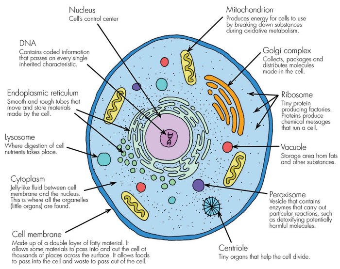
Animal cell parts and functions coloring sheet – This coloring sheet, designed for educational purposes, will leverage visual appeal to enhance understanding of animal cell structures. The layout prioritizes clarity and avoids overwhelming the user with excessive detail, focusing instead on the key organelles and their relative positions within the cell. The color scheme is carefully considered to facilitate differentiation and memorization.The design aims to present a simplified, yet accurate, representation of a typical animal cell, suitable for a younger audience while still retaining scientific accuracy.
This approach acknowledges the limitations of a coloring sheet medium while maximizing its pedagogical potential.
Organelle Placement and Visual Hierarchy
The cell membrane will form the outer boundary of the sheet, a large, rounded shape providing a clear frame for the internal structures. The nucleus, being the largest organelle, will be centrally located, establishing a visual anchor point. Other organelles will be strategically placed around the nucleus, reflecting their relative positions within a real cell, to promote spatial understanding.
Smaller organelles, like ribosomes, will be grouped together for clarity rather than scattered randomly, avoiding visual clutter. The Golgi apparatus and endoplasmic reticulum will be depicted as interconnected networks, showcasing their functional relationship. This organization enhances visual coherence and reinforces the interconnected nature of the cell’s machinery.
Organelle Labeling and Font Selection
Each organelle will be clearly labeled using a sans-serif font, such as Arial or Calibri, in a size easily readable by the target age group. Labels will be placed adjacent to the corresponding organelle, with connecting lines to avoid ambiguity. The font size will be proportionally scaled to the size of each organelle, ensuring readability without visual interference. For instance, the label for the nucleus will be larger than the label for a lysosome, reflecting the relative size difference.
The font style will be consistent throughout the sheet for a professional and uncluttered look.
Color Scheme for Organelle Differentiation
The color scheme will employ distinct and easily distinguishable colors for each organelle. This will aid memorization and reinforce the individual functions of each component. For example, the nucleus might be colored a muted purple to represent its role as the control center. Mitochondria, the powerhouses of the cell, could be a vibrant red to reflect their energy-producing function.
Yo, digging that animal cell parts and functions coloring sheet? It’s like, totally educational, right? But if you’re feeling the need for a lil’ break from organelles, check out these free coloring pages of animals for a quick creative boost. Then, get back to mastering those cell structures – mitochondria are totally the powerhouses, you know!
The endoplasmic reticulum could be a light blue, while the Golgi apparatus could be a slightly darker shade of blue to highlight their relationship. Lysosomes might be a deep orange, and ribosomes a light green. This approach will not only enhance visual appeal but also facilitate learning through color-coding. The color palette will be selected to avoid any potential color blindness issues.
Educational Applications of the Coloring Sheet
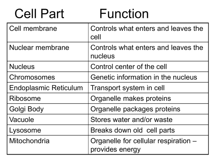
This coloring sheet, depicting the components of an animal cell, transcends mere entertainment; it serves as a potent tool for reinforcing biological concepts in students of varying ages and learning styles. Its effectiveness stems from its ability to engage students visually and kinesthetically, promoting deeper understanding and retention compared to traditional rote learning methods. The visual nature of the activity caters to diverse learning preferences, offering a valuable alternative for students who struggle with abstract concepts.The coloring sheet facilitates a multi-sensory learning experience.
Students not only passively receive information but actively participate in the learning process by coloring and labeling cell components. This active engagement significantly improves memory retention and comprehension. Furthermore, the act of coloring encourages focus and concentration, essential for effective learning.
Lesson Plan Integration
The coloring sheet can be seamlessly integrated into various lesson plans. For instance, it can be used as a pre-activity to introduce the topic of animal cells, generating student interest and providing a visual framework for subsequent lectures or discussions. Following the coloring activity, teachers can lead a class discussion, prompting students to describe the functions of each labeled component.
This can be followed by more advanced activities, such as creating models of animal cells using readily available materials, further solidifying their understanding. The coloring sheet could also serve as a post-activity, reinforcing concepts learned and allowing students to self-assess their comprehension. For instance, a quiz could follow the coloring activity, testing their knowledge of cell structures and functions.
Memorization of Animal Cell Components
The coloring sheet’s effectiveness in memorization stems from its visual and interactive nature. By associating each cell component with a specific color and label, students create strong visual-spatial connections in their brains. This method taps into visual learning preferences, leading to improved recall. Repeated engagement with the coloring sheet, perhaps as a review tool, further reinforces these connections, enhancing long-term retention.
The act of labeling each component further strengthens memory by associating the visual representation with the written name. This dual-sensory approach is significantly more effective than simply reading a textbook description.
Adaptability for Different Age Groups
The coloring sheet’s adaptability lies in its simplicity and versatility. For younger students (elementary school), the focus can be on basic cell components and their general functions, using simpler terminology and brighter colors. Older students (middle and high school) can tackle more complex structures and detailed functions, incorporating more challenging vocabulary and requiring them to research additional information about each component.
Furthermore, the sheet can be adapted to incorporate different learning styles. For example, kinesthetic learners could be encouraged to create three-dimensional models of the cell after completing the coloring sheet, while auditory learners could benefit from a teacher-led discussion focusing on the cell’s functions. The sheet can also be modified to incorporate different levels of difficulty, from simple coloring to complex labeling and detailed descriptions.
For instance, a more challenging version might require students to identify the functions of each organelle without labels, encouraging deeper comprehension.
Advanced Concepts and Extensions
This section delves into more complex aspects of animal cell biology, moving beyond the basic organelles to explore the intricate mechanisms that govern cell structure, function, and reproduction. A superficial understanding of cell components is insufficient; a critical examination of their interactions and roles is necessary for a complete picture. The following discussion aims to provide that deeper, more nuanced perspective.
Cytoskeleton’s Role in Cell Shape and Movement, Animal cell parts and functions coloring sheet
The cytoskeleton, a dynamic network of protein filaments, is far more than mere scaffolding. It’s a highly regulated system crucial for maintaining cell shape, facilitating intracellular transport, and enabling cell motility. Microtubules, intermediate filaments, and microfilaments—the three main components—interact in a complex interplay to achieve these functions. For instance, microtubules, the largest filaments, form tracks for motor proteins like kinesin and dynein to transport organelles and vesicles.
Microfilaments, composed of actin, are vital for cell crawling and muscle contraction. Disruptions to the cytoskeleton, often caused by genetic mutations or toxins, can lead to significant cellular dysfunction and disease. Consider the debilitating effects of certain cancers, where uncontrolled cell division and migration are often linked to cytoskeletal abnormalities.
Centrioles’ Role in Cell Division
Centrioles, cylindrical structures composed of microtubules, play a pivotal role in cell division. They organize the microtubules into the mitotic spindle, a crucial apparatus that separates chromosomes during mitosis and meiosis. The precise alignment and segregation of chromosomes are essential for generating genetically identical daughter cells. Failure in this process can result in aneuploidy, a condition characterized by an abnormal number of chromosomes, often leading to developmental disorders or cancer.
The centrioles’ function highlights the critical link between cell structure and the accurate transmission of genetic information.
Differences Between Animal and Plant Cells
Understanding the distinctions between animal and plant cells is fundamental to grasping the diversity of life. While both are eukaryotic cells sharing some basic organelles, significant differences exist that reflect their unique adaptations.
- Cell Wall: Plant cells possess a rigid cell wall made of cellulose, providing structural support and protection, absent in animal cells.
- Chloroplasts: Plant cells contain chloroplasts, the sites of photosynthesis, enabling them to produce their own food; animal cells lack this capacity.
- Vacuoles: Plant cells typically have a large central vacuole for storage and turgor pressure regulation; animal cells have smaller, more numerous vacuoles.
- Plasmodesmata: Plant cells are interconnected by plasmodesmata, channels that allow communication and transport between adjacent cells; animal cells lack these structures.
- Centrioles: While most animal cells possess centrioles, plant cells generally lack them, utilizing other mechanisms for spindle organization during cell division.
Examples of Specialized Animal Cells and Their Unique Adaptations
The remarkable diversity of animal life is reflected in the specialized cells that perform specific functions. Consider the following examples:
- Neurons: These cells, with their long axons and dendrites, are highly specialized for rapid signal transmission throughout the nervous system. Their elongated shape and intricate branching patterns are crucial for their function in communication and coordination.
- Muscle Cells: Muscle cells, packed with contractile proteins like actin and myosin, are designed for generating force and movement. Their elongated, fiber-like structure and specialized protein arrangements enable efficient contraction and relaxation.
- Red Blood Cells: Erythrocytes, or red blood cells, are highly specialized for oxygen transport. Their biconcave disc shape maximizes surface area for efficient gas exchange, and they lack a nucleus to accommodate more hemoglobin.

